

Finding a lump doesn't necessarily signify a severe health issue, and even if it turns out to be a type of cancer, there could be several treatment approaches available.
Explaining the terminology
The language used to describe such conditions can be confusing, and you might come across words like lump, mass, growth, tumour, neoplasia or cancer, often used interchangeably.
Generally, lump and mass are descriptive terms used when the exact nature of the issue isn't clear yet.
A lump could be a fluid filled cyst, an abscess, a result of inflammation, perhaps around a hair follicle or due to a foreign body such as a thorn, or a mass originating from fat, muscle or bone.
This growth, mass or tumour can either be benign, indicating that it's non-cancerous and does not spread elsewhere in the body.
Or it could be malignant, meaning that it's cancerous and likely to spread to other parts of the body.
Consultation
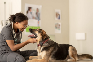
We would advise booking an appointment with the vet so they can examine the lump.
We cannot tell what a mass is just by looking at it but your vet will examine the following aspects:
It helps reduce Is the lump firm or soft?
Is it mobile and 'loose' within the skin layer, or is it attached to deeper structures?
Does it cause pain?
Has there been any hair loss over the lump, or has it become red, sore or ulcerated?
Sampling the lump
The objective of sampling is to understand the nature of the lump, which informs the vet's strategy for its treatment.
When we take a sample, we're trying to answer questions such as:
- Is it inflammation or growth?
- If it's a tumour, what kind is it and is it benign or malignant?
- What is the best course of action?
- Planning - can the lump be excised locally or do we need to remove larger amounts of surrounding tissue to avoid recurrence?
How will the vet decide which type of sample to take?
Several factors will influence this decision, including the mass's location, size, your pet's temperament and the position of the mass on the body.
- Fine needle aspirate (FNA) sampling technique: A small needle, like those used for blood samples, is inserted into the mass, and sometimes suction is applied with a syringe to increase the chance of yielding a good number of cells.
After removing the needle, the contents from the needle hub are transferred onto a microscope slide.
This is then analysed in-house or sent to an external lab.
This method is minimally invasive and can typically be carried out during your consultation or scheduled for a longer appointment.
Your pet usually doesn't require any sedation/anaesthetic if they are co-operative and the same can be taken safely.
It is relatively affordable compared to a biopsy, and results are typically available within a few days.
Whilst most of the time the lab can provide an answer, since this method collects only a few cells and not a tissue section, there is always a chance that we may not get a diagnostic sample and definitive answer.
Therefore we may need to contemplate
- 'plan B' - a biopsy. Biopsy: The alternative method is a biopsy, which involves extracting a piece of tissue for laboratory histopathology analysis.
This requires a surgical procedure, meaning your pet would need to be admitted for the day to undergo the operation under sedation or general anaesthesia.
The benefit of this method is that it provides the lab with more tissue to examine, reducing the likelihood of an inconclusive result.
However, it does require anaesthetic and is consequently more costly.
Typically, a biopsy does not completely remove the lump (this is an incisional biopsy), implying a potential need for a second surgery.
Why not just remove the mass?
We usually recommend sampling the lump initially.
This enables the vet to identify the nature of the lump which in turn helps us to recommend the most effective treatment course.
It helps reduce uncertainty and the chances of recurrence and complications.
However, in certain situations, some exceptions might be necessary, so we suggest discussing these options with us.
In some instances, if the mass is extremely small and located where there is ample 'extra' skin, a biopsy may lead to complete excision.
But as mentioned earlier, if the histopathology results indicate that the mass has infiltrated the surrounding tissues, a repeat surgery may be required to remove additional skin.
To book in for your pet in for a health check or testing, please call Claro Hill Vets on 01423 228080 or visit www.clarohillvets.co.uk.